Medical Images Comparison
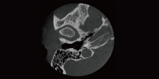
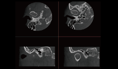
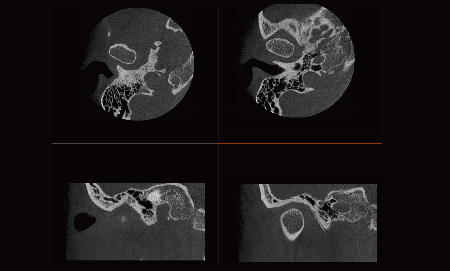
By loading multiple images, it is possible to perform single-slice comparison medical imaging. It can also compare multiple slices simultaneously by adjusting the layout view, as well as set multi-sequence synchronous page turning, zooming, panning, and adjusting window width and window level, thereby saving the comparison image function.
CPR-Surface Reconstruction
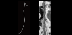
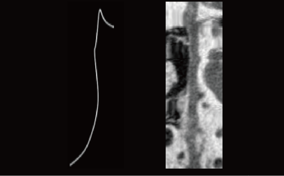
Precisely identify the centerline of the tubular structure based on a semi-automatic method. Visualize 3D curved tubular structures with advanced resampling algorithms to 2D image sequences, enabling intuitive inspection of tubular structures.
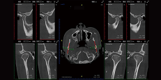
TMJ Diagnosis
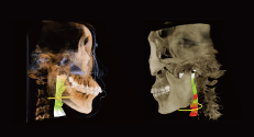
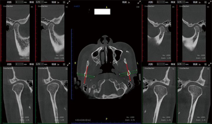
SmartV-ENT has a visual pattern of comparison of left and right joints, allowing doctors to evaluate the diagnosis and treatment effect of temporomandibular joint diseases.
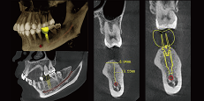
Implant Simulated
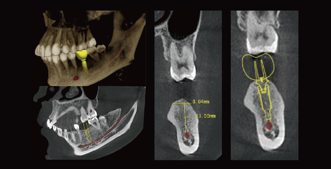
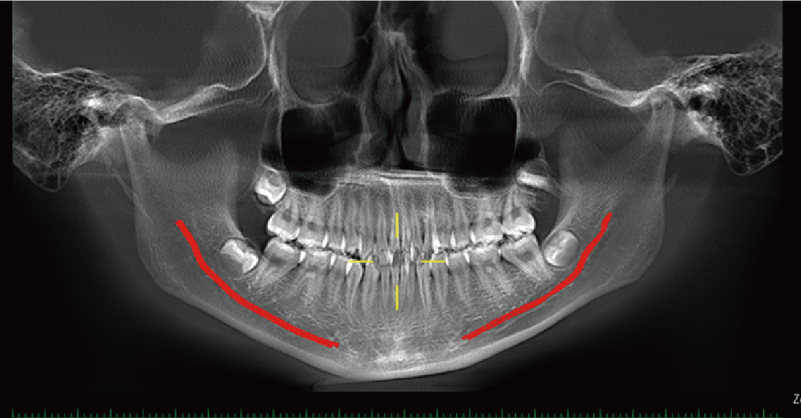
Neural tubes will be highlighted, which presents a relationship between the location of the implant and optimal length. This is the best way to improve the success rate for implant surgery.
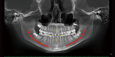
Neural Tube Automatic
Labeling
Label the neural tube automatically in the CT image, providing great convenience for diagnosis.
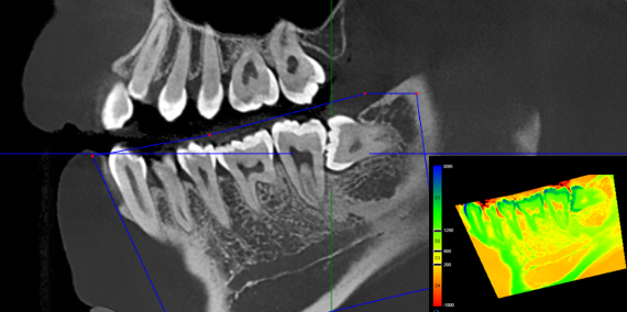
Regional Statistics
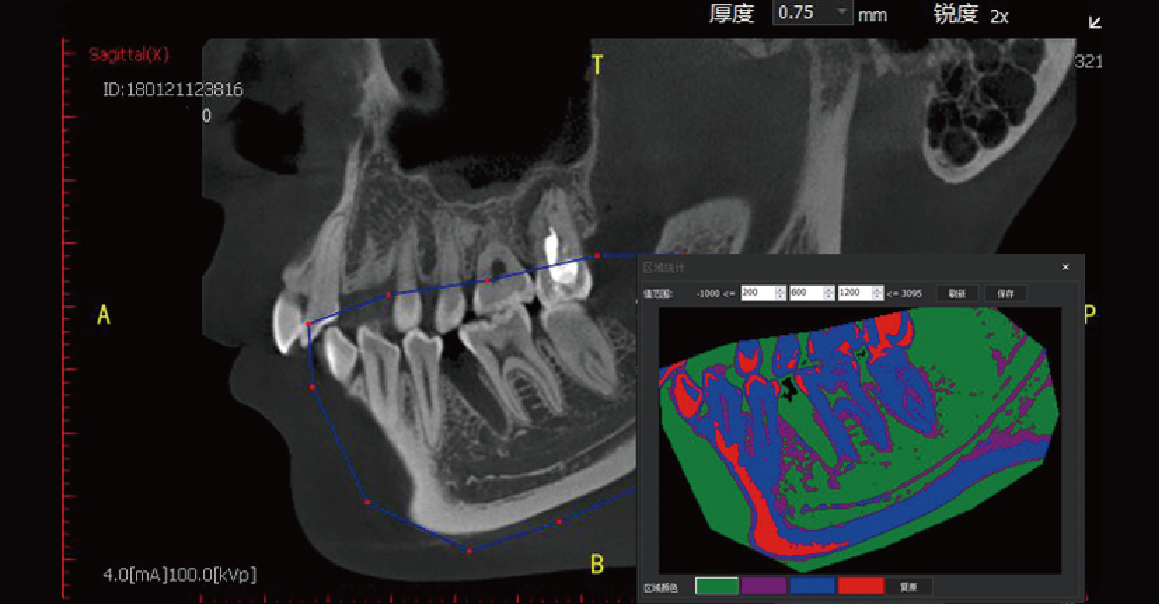
Used to evaluate bone mineral density in a selected area.
 English
  Русский
 Français
 Español
